EVO

Modular SEM Platform for Intuitive Operation, Routine Investigations and Research Applications

The instruments of the EVO family combine high performance scanning electron microscopy with an intuitive, user-friendly experience that appeals to both trained microscopists and new users. With its comprehensive range of available options, EVO can be tailored precisely to your requirements, whether you are in life sciences, material sciences, or routine industrial quality assurance and failure analysis.

  • Versatile solution for central microscopy facilities or industrial quality assurance laboratories
  • Excellent images from any real-world sample
  • Maximum image quality with the lanthanum hexaboride (LaB6) emitter
  • Imaging and analytical excellence on non-conductive and uncoated samples
  • Workflow automation and data integrity

Highlights

Class-Leading Usability

SmartSEM Touch puts int
eractive workflow control directly at your fingertips. It is quick and easy to learn, dramatically reducing training effort
and costs. Within minutes, even new users will begin capturing stunning images. This user interface also supports industrial
operators who require automated workflows for repeatable inspection tasks.

Embryonic stage of a squid

Excellent Image Quality

EVO excels at extracting the maximum data quality from uncoated and unaltered samples. EVO also safeguards data quality on hydrated and heavily contaminated samples, by allowing these samples to remain in their native state. Additionally, the LaB6 emitter will give that extra bit of resolution, contrast and signal-to-noise that is important when imaging and microanalysis get challenging.

EVO Plays Well with Others

EVO can be configured to be part of a semi-automated multi-modal workflow, with tools for seamless relocation of regions of interest and integrity of data collected from multiple modalities. Combine light and electron microscope data for material characterization or parts inspection. Or combine EVO with ZEISS light microscopes for correlative particle analysis.

Embryonic stage of a squid

Get More Hands on Deck

Depending on the actual laboratory environment, operation of the SEM can be the exclusive domain of expert electron microscopists. But this situation is challenged by the very common necessity that non-expert users, such as students, trainees, or quality engineers, also require data from the SEM. EVO takes both requirements into account, with user interface options that cater to the operational needs of experienced microscopists as well as non-micoscopists.

Navigation Camera

  • A camera can be mounted either to the chamber to monitor the position of the samples relative to the pole piece mounted backscattered detector (chamberscope); or on the vacuum chamber door (navigation camera) to enable a helicopter view of the arrangement of samples or parts on the sample holder. This view can then be used to set up predefined locations of interest identified from a light microscope image, and for easy navigation during the entire sample investigation process.

  •  

PCB - Annular Light

Coaxial epi-Illumination S Particularly Suitable for Flat, Specular Specimens​

Automated Intelligent Imaging

EVO enables automated, unattended acquisition of images across sample batches. ZEISS Automated Intelligent Imaging is perfectly suited to routine inspection. It enables the user to define a boundary region, automatically generate regions of interest determined by the required field of view or magnification, and begin automated acquisition. Automated Intelligent Imaging will improve your sample throughput, boosting productivity and performance.

Take Your Investigation to the Next Level

  • Better Data with a Lanthanum Hexaboride (LaB6) Electron Emitter

    Electron emission from a lanthanum hexaboride cathode, rather than a traditional tungsten hairpin filament, provides the reassurance that every extra bit of image quality is there when you need it. And that is a benefit you can put into action in two ways:

    • At equivalent electron probe sizes (i.e. resolution), there is more probe current to work with, which makes image navigation and optimization much easier.
    • At equivalent probe currents (signal-to-noise), the beam diameter is much smaller, resulting in enhanced image resolution.
evo-lab6-cathodeevo-tungsten-filament

EVO Plays Well with Others

Expand Your Possibilities with ZEISS ZEN core

Your Software Suite for Connected Microscopy and Image Analysis

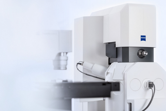
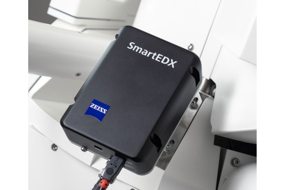
With ZEISS being the supplier of microscopy and metrology systems, you can expect EVO to play extremely well with other ZEISS solutions. Establish a highly-productive multi-modal workflow between (digital) light microscopes and EVO. Combine the unique optical contrast methods of your light microscope with the equally unique imaging and analytical methods of your SEM to obtain complementary data, and hence more meaningful information about the material, quality or failure mechanism of your sample.

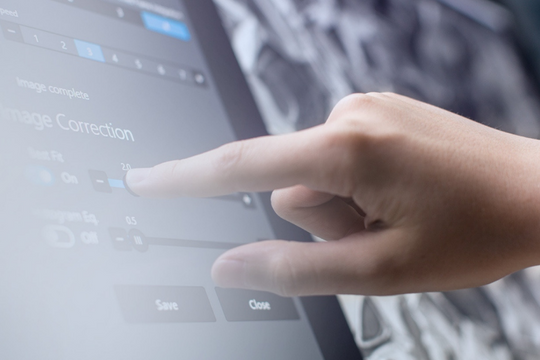
Take advantage of ZEN core as your hub for connected microscopy. Customize its functions to your specific applications and define workflows that consider the experience level of the  microscopists in your multi-user environment.

Enjoy its highlights:

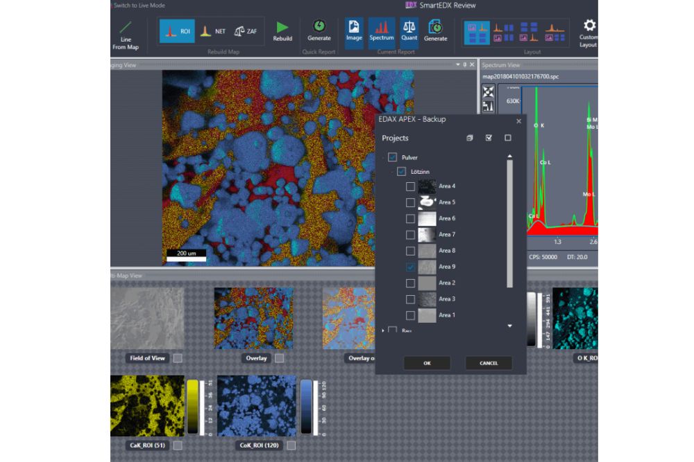
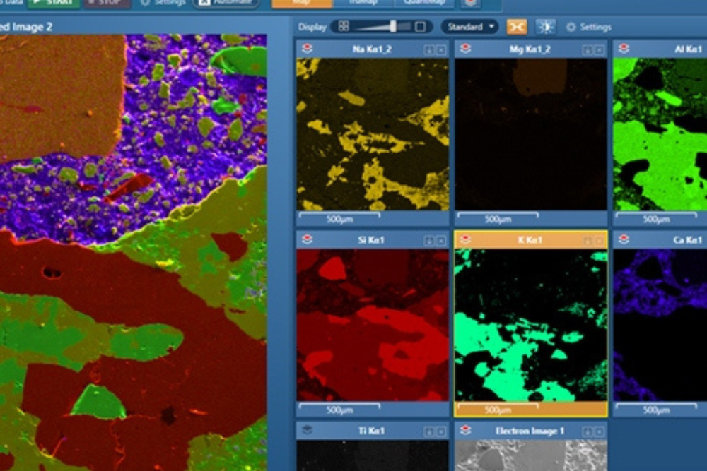
  • Correlative Microscopy: Sample and data exchange between light, digital, and electron microscopes
  • Contextual Data Representation: Data visualization and organization across scales and imaging modalities
  • Metallographic Applications incl. Microsoft Word-based Reporting: Integrated reporting across connected images and datasets
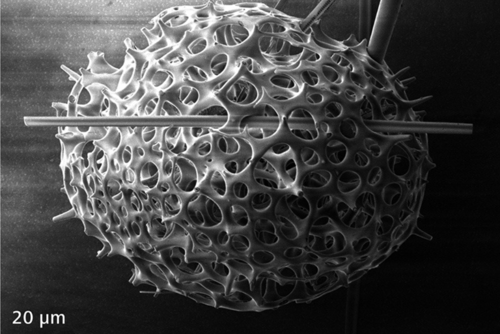
  • Automated Image Analysis: based on deep learning: Image segmentation based on machine learning algorithms.

EDX Solutions for Microanalysis Applications


Workflow-Guided Graphical User Interface

SmartSEM Touch puts int
eractive workflow control directly at your fingertips. It is quick and easy to learn, dramatically reducing training effort
and costs. Within minutes, even new users will begin capturing stunning images. This user interface also supports industrial
operators who require automated workflows for repeatable inspection tasks.

Optimized for Routine Microanalysis Applications

SEM and EDS have to be paired with careful consideration. SmartEDX on EVO is ideally suited for routine microanalysis applications, particularly for customers with high standards for data reproducibility. It provides highest throughput at 129 eV energy resolution and 1-5 nA probe current – typical EVO operating condition. SmartEDX is optimized to detect low energy X-rays from light elements thanks to superior transmissivity of the silicon nitride window.

Choose between Different EDX Detector Configurations

The single PC solution offers you various EDS configurations: the Xplore 15, 30 and the Ultim Max 40 detectors from Oxford Instruments can be ordered.  

Simplify Operation and Collect EDS Data More Efficiently

Control both EDS and SEM in parallel using one single PC. This integration improves usability. At the same time, you will enjoy dedicated user interfaces for your microscope and your EDS system. Reduce your EDS acquisition time by leveraging the optimized detector integration that boosts the EDS signal inputs by at least 17%.

Total ZEISS Service and System Support

Because SmartEDX is supported entirely by ZEISS, this EDS solution is ideal for customers with a vested interest in streamlining their number of analytical equipment suppliers. All installation, preventive maintenance, warranty, diagnostics and repair, spare part logistics, and inclusion in total system service contracts are fully handled by ZEISS, making support of your analytical SEM solution easy.

The EVO Family

ZEISS EVO 10

Choose EVO 10—with optional backscatter detector and Element EDS system—to be your entry point to scanning electron microscopy, at a remarkably affordable price. Even this smallest of EVO vacuum chambers is well differentiated from tabletop SEMs. Your investment in EVO now assures that you are ready for applications that require more space and ports than you anticipate today.

ZEISS EVO 15

EVO 15 demonstrates the flexibility concept of the EVO family and excels in analytical applications. Opt for the larger vacuum chamber of the EVO 15, and add variable pressure for imaging and analysis of non-conductive samples or parts, and you have a versatile, multi-purpose solution for central microscopy facilities or industrial quality assurance laboratories.

ZEISS EVO 25

EVO 25 is the industrial workhorse solution with enough space to accommodate even the largest parts and assemblies. Expand EVO 25 capabilities further with an optional 80 mm Z travel stage that can handle weights up to 2 kg even with tilt. Additionally, the large chamber will accommodate multiple analytical detectors for the most demanding microanalysis applications.

Accessories

Beam Deceleration Imaging

Use beam deceleration imaging to investigate especially delicate specimens. Gain improved image quality and minimize sample damage. Image non-conducting specimens with higher resolution, more surface sensitivity, and more contrast. A bias voltage is applied to your sample. This reduces the effective landing energy on your sample while the primary energy is kept high.

Applications

Zinc-phosphate E-coating, imaged with SE detector in high vacuum.

Car seat cushion foam, imaged uncoated in Variable Pressure mode with the BSE detector.

Stainless steel fracture surface, imaged with secondary electrons in high vacuum.

Similar Products

Similar Products

Similar Products

Let’s find solution

to your TECHNICAL PROBLEMS.

Get In Touch